Research

Research Topics

The Pelletier laboratory studies several facets of centrosome biogenesis and function, with a particular emphasis on how their perturbation can lead to devastating developmental diseases and cancer. Centrosomes are composed of a centriole pair surrounded by a proteinaceous matrix composed of over 100 proteins termed pericentriolar material (PCM). By organizing PCM, centrosomes orchestrate several fundamental cellular processes including bipolar mitotic spindle assembly, which plays a determining role in the faithful and accurate segregation of chromosomes to progeny. Centrosome also template the formation of cilia and flagella, key cellular organelles, that play important roles during animal development, cell motility and signaling pathways. Using functional genomic approaches, in combination with cutting-edge microscopy, biochemistry, proteomics and advanced single-cell analytics the overreaching goal of our lab is to identify and study novel proteins required for proper centrosome-cilia biogenesis and function and to investigate their potential roles in human development and disease.

Research in the Pelletier lab revolves around the following axes:

project1_clip_image003f

The centrosome is the major cellular microtubule-organizing center (MTOC) in mammalian cells. Centrosomes are composed of a pair of barrel-shaped structures called centrioles, surrounded by a proteinaceous matrix termed pericentriolar material (PCM). Centrioles mark the site at which PCM is recruited and hence, through the control of their duplication, define the number of centrosomes present. Prior to mitosis, duplicated centrosomes need to increase sufficiently in size by recruiting more PCM leading to an increase in MT nucleation capacity, which is needed to orchestrate the assembly of a robust bipolar spindle. Centrioles have fascinated biologists ever since their discovery by Boveri in 1888, not only for their stunning 9-fold symmetry but also for their enigmatic mode of duplication and assembly. We initially defined using the nematode C. elegans as a model system, a structural and molecular pathway that regulates centriole duplication (Pelletier et al., Nature 2006). This core duplication pathway is conserved in human cells and is a focus of study in the laboratory as well as the identification of novel regulators of centriole duplication. Another keen interest of the laboratory is to understand how centrosome assemble and increase in size and microtubule nucleation capacity at the onset of mitosis. Using sub-diffraction resolution imaging, we have defined higher-order organizational properties of pericentriolar material and centriole (Lawo et al., Nature Cell Biology, 2012 and Comartin et al., Current Biology, 2013).  Thus, an important goal of the laboratory is to decipher the cellular pathways that regulate centrosome duplication and assembly, to identify novel regulators of these processes and to delineate how these novel molecules contribute to centrosome biogenesis. We have recently identified a number of novel regulators of centrosome function through proximity mapping and are currently studying them in greater detail (Gupta et al., Cell, 2015). For more information on the higher-order organizational properties of centrosomes, see our review on this topic (Mennella et al., Trends in Cell Biology, 2014).

Pelletier et al. Nature 2006
Lawo et al. Nature Cell Biology 2012
Comartin et al. Current Biology 2013
Gupta et al. Cell 2015
Mennella et al. Trends in Cell Biology 2014

project2_clip_image002f

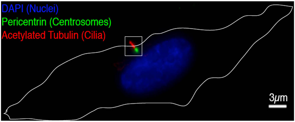
Cilia are microtubule-based structures that protrude from the cell surface and participate in a plethora of developmental processes and signaling events. Disrupting their assembly and function can lead to several diseases including myriad ciliopathies. Although motile and sensory cilia are without a doubt most famous for their paramount roles in cell motility, hearing, and in cleansing the respiratory track of pathogens, it has become increasingly clear that most cell types have immotile primordial cilia referred to as primary cilia. Primary cilia are implicated in key developmental signaling events mediated by the Wnt and Hedgehog (Hh) morphogens, planar cell polarity (PCP) and are defective in signaling events leading to the onset of disease. It is therefore critical that we understand the composition of cilia and the mechanisms underpinning their assembly and how they efficiently transduce extracellular cues through various signaling pathways. Using proximity-dependent protein identification (BioID) we have recently defined a dynamic proximity interaction landscape of the human centrosome-cilium interface (Gupta et al., Cell, 2015) highlighting vast and novel functional interaction space in those areas that remains virtually unexplored. Thus, a major goal of the laboratory is leverage this information to understand the molecular mechanisms that govern cilia formation and their roles in diverse signaling pathways and the onset of human ciliopathies. For more on this topic, see our current review (Goncalves et al., Mol. Cells, 2017).

Gupta et al. Cell 2015
Goncalves et al Mol Cells 2017


project3_clip_image002f

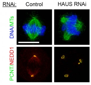
The assembly of a robust mitotic spindle apparatus, capable of accurately segregating chromosomes to progeny cells, requires the concerted action of several intricate cellular pathways. These include regulators of microtubule dynamics, centrosome biogenesis, kinetochores assembly, molecular motors, non-motor spindle proteins and chromosome-based microtubule nucleation. Although several key players in each of these pathways have been identified and studied with some detail, we have only begun to understand the relationship between these proteins and critically, several key regulators arguably remain to be identified. In the Pelletier lab we use high-content / high-resolution imaging and screening approaches to probe the human genome to identify and study novel regulators of mitotic spindle assembly and delineate their interplay with the various processes enumerated above. A major ongoing effort in the laboratory is to use high-content screening approaches to identify novel regulators of mitotic spindle assembly. Such an approach has recently led to the identification of a novel eight-subunit complex called HAUS required for centrosome and mitotic spindle integrity, which shares homology to Drosophila melanogaster Augmin (Lawo et al, Current Biology, 2009) and more recently, in collaboration with the Gingras and Raught laboratories a functional proteomics screen to identify novel phosphatases required for normal cell cycle and mitotic progression (St-Denis et al., Mol Cell Prot, 2015 and St-Denis et al., Cell Reports, 2016). This serves to exemplify how functional genomics can be used to identify and study novel protein complexes that play instrumental roles in mitotic spindle assembly. For a comprehensive review on the topic of mitotic spindle assembly, have a look at our recent review in Nature Reviews in Molecular Cell Biology (Prosser et al., Nat Rev Mol Cell Biol 2017).

Lawo et al, Current Biology 2009
St-Denis et al. Mol Cell Proteomics 2015
St-Denis et al. Cell Reports 2016
Prosser et al. Nat Rev Mol Cell Biology 2017

In collaboration with the Wrana lab we are interested in understanding the role of centrosomes in the regulation of cancer cell motility, migration and metastasis. Together our groups have identified a role for key centrosome components in exosome-mediated breast cancer cell motility and protrusion formation. Using this system, they have begun to probe the proximity interaction landscape of centrosome/cell polarity proteins to identify which protein kinases are involved in this process. In pursuing this approach, they have identified discrete centrosome modules that interact extensively with Wnt-PCP pathway components to drive cell motility. We have demonstrated that, discrete centrosome modules are actively engaged in extensive crosstalk with Wnt-PCP pathway components at the leading edge of cells to drive cell motility through reorganization of actin filaments. Future lines of investigation will revolve around the delineation of interplay between other key signaling pathways, the centrosome-cilium interface and cytoskeletal systems and how this system is controlled during tissue formation, degeneration and regeneration using organoid and mouse models.